


Entertainment

Made In Chelsea Star Tabitha Willett Welcomes Baby Boy Seven Weeks After Wedding
Tabitha Willett, star of Made In Chelsea, has given birth to a baby boy named Orlando, just seven weeks after marrying Harry Hoare at Chelsea Town Hall.


Politics

Trump Furious as Judge Orders His Name Removed from Kennedy Center
Donald Trump slammed a judge's decision to remove his name from the Kennedy Center, calling the venue's path a 'hopeless journey into never never land' after the ruling.


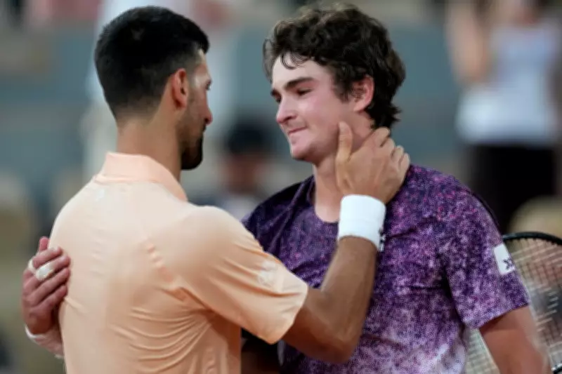
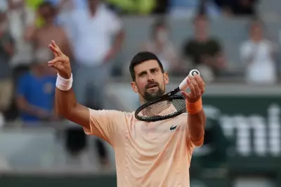
Sports

Arsenal's David Raya Unrecognisable After Horror Facial Injury Transformation
Arsenal goalkeeper David Raya looks completely different after a sickening facial injury in 2018. The Spaniard, now a Premier League champion, underwent reconstructive surgery and has since become a key player for the Gunners ahead of the Champions League


Crime

Teen Charged with Murder of Boy, 15, in London Stabbing
A 16-year-old has been charged with murder after Brayan David Saldarriaga, 15, was fatally stabbed in Hackney, London on 25 May.


Health


Environment

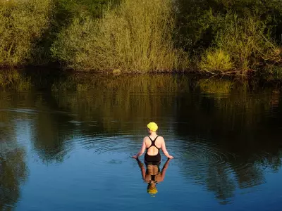
Cheap Homemade Air-Con and Summer Survival Tips
Readers share cheap air-con hacks, external shutter alternatives, and screen time benefits, plus a correction on Southgate's penalty.
Rare Giant Otter Pups' First Swim at Chester Zoo
Watch 15-week-old giant otter triplets Uca, Yali, and Yara take their first swimming lesson with parents Bonita and Manu at Chester Zoo, part of a conservation breeding programme.
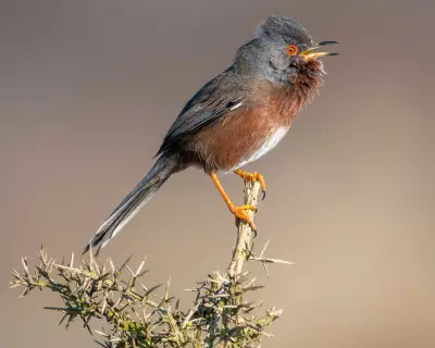
Dartford Warbler Comeback 60 Years After Near Extinction
The Dartford warbler, once on the brink of extinction in England, has seen a 44% increase on RSPB reserves thanks to heathland restoration efforts.
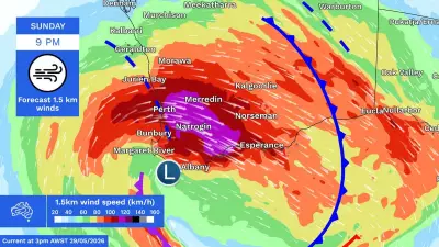
Cyclone-Strength Storm Threatens Western Australia
A major storm system brings cyclone-strength winds to WA, with gusts over 125km/h, heavy rain, and flood risks. The system will move east, bringing icy weather to NSW, Victoria, and SA.
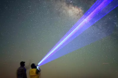
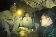
UAE's Darkest Desert Reveals Stunning Milky Way Views
In the UAE's Al Quaa Desert, stargazers escape light pollution to see the Milky Way, reconnecting with the night sky as Bedouins once did.
UK Geography Weekend Special
Weekend special: 100 questions about UK geography. Perfect for students preparing for exams or anyone who loves learning about Britain's diverse landscapes.