
Groundbreaking 3D Mapping of Clitoral Nerves Completed
In a landmark scientific achievement, researchers have successfully mapped the full network of nerves within the clitoris for the first time, using advanced 3D scanning techniques. This breakthrough comes nearly three decades after similar mapping was accomplished for the penis, highlighting a long-standing gap in anatomical knowledge.
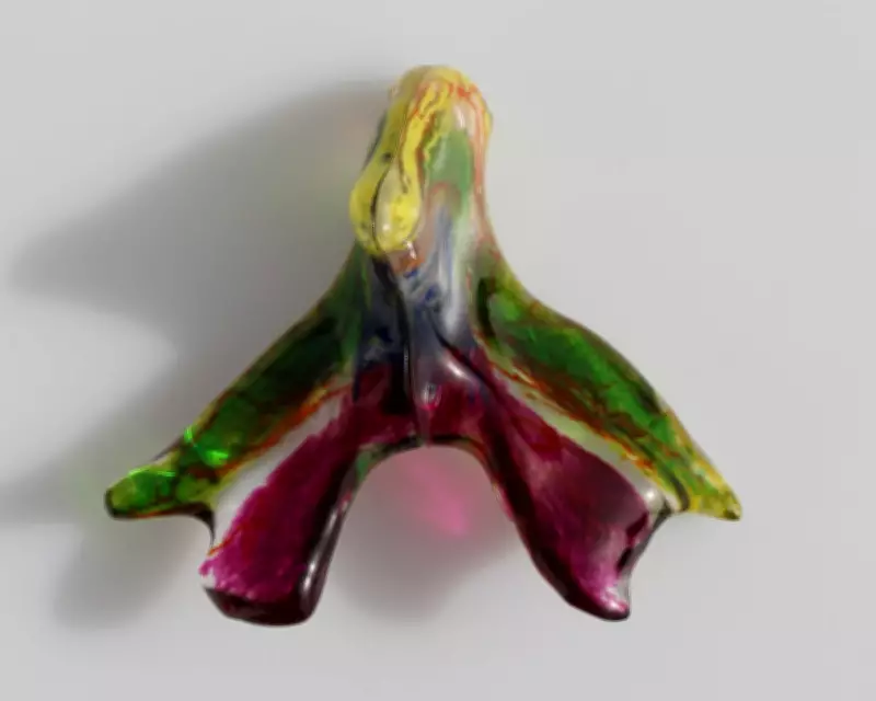
Unprecedented Detail Reveals Anatomical Errors
The study, led by Ju Young Lee, a research associate at Amsterdam University Medical Center, employed high-energy X-rays to generate detailed 3D scans of two donated female pelvises. These scans unveiled the intricate trajectory of five tree-like branching nerves within the clitoris, with the widest measuring 0.7mm across. The findings, reported on the preprint server bioRxiv, challenge existing medical understanding by showing that some nerves extend further than previously thought, particularly to areas like the mons pubis and clitoral hood.
Helen O'Connell, a pioneering urologist from Melbourne who first mapped the clitoris in 1998, emphasized the significance of this work. She noted that the clitoris has historically been overlooked due to cultural taboos surrounding female sexuality, often being omitted from standard anatomy textbooks or inaccurately described.

Implications for Medical Practice and Women's Health
The new 3D map is expected to have profound implications for various surgical procedures. For instance, it could enhance reconstructive surgery after female genital mutilation, a practice affecting over 230 million women globally, by providing a clearer understanding of nerve pathways to preserve sexual function. Additionally, the research may inform treatments for vulvar cancer, gender reassignment surgeries, and cosmetic procedures like labiaplasty, which have seen a significant rise in recent years.
Georga Longhurst, head of anatomical sciences at St George's, University of London, expressed fascination with the high-resolution images, particularly within the glans clitoris, where terminal nerve branches are typically invisible during dissection. This level of detail is crucial for improving outcomes in pelvic surgeries, potentially reducing the decline in orgasmic experience reported by some patients post-operation.
Future Directions and Educational Initiatives
Lee and her team hope to expand public knowledge through initiatives like a clitoris exhibition at Amsterdam University Medical Center, inspired by the Vagina Museum in London. This effort aims to combat societal ignorance and promote a better understanding of female anatomy, ultimately contributing to improved health and wellbeing for women worldwide.